Preclinical Imaging Methodology
Medical physics and instrumentation play an important role in our research program. This aspect of our activity is organized into three main areas.
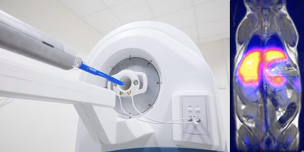
Innovating PET/MRI Technology for Precision Imaging !
Instrumentation
- Development of innovative imaging systems in collaboration with our academic and industrial partners. During IMAPPI, we initiated collaborations with Bioscan, Trifoil Imaging, and MR Solutions Ltd.
- Calibration and fine-tuning of imagers, to obtain high-resolution morphological images (CT, MRI) recalibrated with quantitative functional data (SPECT, PET).
- Reduction of interference between PET and MRI systems.
- Imager performance evaluation based on international standards.
- Design of quality assurance protocols.
- Technology watch to maintain a high level of performance and adaptability to scientific projects.
Preclinical in vivo imaging
- Design of morphological, quantitative and functional imaging procedures to meet the needs of basic and applied research teams.
- Participation in in vivo validation of the use of molecular probes and nanomaterials, compatible in particular with PET/MRI.
Dosimetry of ionizing radiation
- Assessment of absorbed dose resulting from nuclear or radiological examinations.
- Optimization of quantitative data analysis methods.
- Implementation of personalized dosimetry for internal vectorized radiotherapy.
Since 2012, IMATHERa has been home to the Equipex “future investment” program IMAPPI (Integrated Magnetic resonance And Positron emission tomography in Preclinical Imaging), funded by the French National Research Agency (ANR) to the tune of €7.3 million. During the three development phases of the IMAPPI project, a preclinical PET/MRI imaging prototype in which the PET is integrated into the center of the MRI magnet was designed, assembled and validated. This system, whose performance was published in 2021, has since been commercialized and is used by several research teams around the world.
From 2021 onwards, the IMAPPI project will embark on a new phase of research designed to capitalize on the highly complementary nature of PET and MRI data.
Our current projects focus on the following areas, among others :
- improving cardiac imaging, notably for early detection of the cardiotoxic effects of cancer chemotherapy and imaging of ischemic heart disease.
- imaging of the tumor microenvironment and biomarkers of interest in immunotherapy, in particular microcirculation and tissue oxygenation.
- integrating radiomics and artificial intelligence into projects.

Associated publications:
Vrigneaud J.M., McGrath J., Courteau A., Pegg R., Gomis A.S.-P., Camacho A., Martin G., Schramm N., Brunotte F. Initial performance evaluation of a preclinical PET scanner available as a clip-on assembly in a sequential PET/MRI system. 2018. Phys. Med. Biol. 63, 125007.
S. Bricq, H. L. Kidane, J. Zavala-Bojorquez, A. Oudot, J. M. Vrigneaud, F. Brunotte, P. M. Walker, A. Cochet, A. Lalande, Automatic deformable PET/MRI registration for preclinical studies based on B-splines and non-linear intensity transformation. Med Biol Eng Comput 56, 1531-1539 (2018).
Vrigneaud J.M., Walker P., Barbier B., Camacho A., Oudot A., Collin B. Brunotte F. Performance evaluation of the PET component of a sequential APD-based micro-PET/MR imaging system. 2017. Biomed. Phys. Eng. Express 3, 035006.
Pierre-Marc Jodoin, Fredy Pinheiro, Alexandra Oudot, Alain Lalande. Left-Ventricle Segmentation of SPECT Images of Rats. 2015. IEEE Transactions on Biomedical Engineering, Institute of Electrical and Electronics Engineers, 62(9), pp. 2260–2268.
Brunotte F., Haas H., Collin B., Oudot A., Bricq S., Lalande A., Tizon X., Vrigneaud J.M., Walker P. Integrated PET/MRI in preclinical studies State of the art. Tijdschrift voor nucleaire geneeskunde, 2013, 35